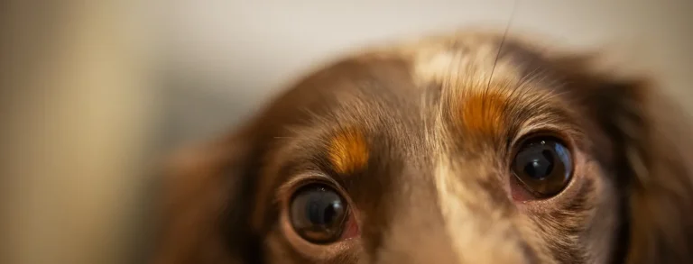
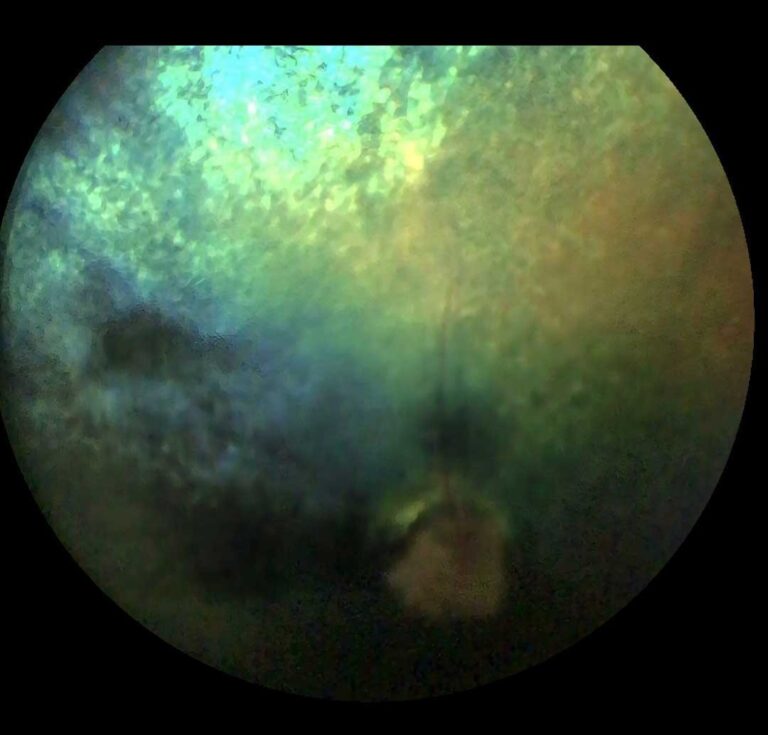
A cataract is a clouding of the lens, within the eye . As the cataract progresses (matures) the opacity turns from blue to grey , and ultimately white . Once the lens has turned white the cataract is referred to as ‘mature’ . At this point it is very unlikely that the animal will be able to see out of this eye.
We will be able to tell if the eye has a true cataract, or just ageing changes of the lens, which will cause a blue discolouration of the lens.
Cataracts can form for many reasons but normally the patients we see either have inherited cataracts (a genetic fault) , or they have had inflammation in their eye and a secondary cataract , or they are Diabetic . 70% of Diabetic dogs will develop a cataract within a year of onset of the Diabetes . The faster the cataract develops the more likely it is to cause secondary problems and therefore the quicker it should be removed . This is especially true in young or diabetic patients .
How do we remove a cataract?
Cataract surgery in animals is done using Phacoemulsification (Phaco) , which is the same technique used in humans . This involves creating a small incision at the edge of the clear surface of the eye (cornea) . A second incision is then made in the capsule surrounding the lens and the Phaco tip is introduced through these incisions and into the lens . Ultrasonic energy is then passed via the Phaco tip into the lens causing it to shatter into tiny pieces which are immediately removed using suction . Once the cataract has been completely removed a new prosthetic lens is introduced into the capsular bag , allowing the light to be focused back onto the retina, returning vision .Occasionally we cannot introduce a new lens . These animals also regain vision but without the same detail. The operation is completed by suturing the corneal incision and re-inflating the eye . Cataract surgery is approximately 94% successful in dogs and cats . This is not as good as humans because the animal lens is larger and the animal eye is more easily inflamed during the surgery. Very occasionally the surgical complications result in a chronically painful eye which has to be removed at a later date . Thankfully this is rare.
What can go wrong?
Common complications of surgery:
- Uveitis (inflammation of the eye)
- Post operation hypertension (high pressure in the eye) and this can progress to glaucoma
- Mild scarring of the lens capsule that usually never affects the vision
- Corneal Ulcer (usually mild and treated medically)
- Reaction or infection of the corneal sutures (usually mild and treated medically)
Less common/rare:
- Retinal Detachment (total permanent blindness)
- Bleeding in the back of the eye (this can clear but can often lead to reduced vision)
- Movement of the new Intra-ocular lens
Longterm
This vision-saving surgery is hard work in terms of aftercare as lots of eye drops and check-ups are required after surgery, but it means that we can give dogs their sight back.
If you thick your dog or cat could be developing cataracts, we can take a look and discuss the estimated success rate for your particular pet as every animal and every eye is different. We create tailor-made plans for each patient to allow the very best outcomes.